History
X-rays are a form of electromagnetic radiation. They were discovered accidentally by Wilhelm Conrad Roentgen, a german physicist in 1895. He had been experimenting with radiation. When he saw his wifes hand appear on an X-ray photo, he realized the potential of x-ray imaging in collecting information on patients. He worked hard on his research for two months and then published his results in the proceedings of the Physical Medical Society of Wurzburg on december 18th, 1895. He used the letter X to describe his newfound, unknown rays. Since then they are referred to as X-rays or roentgen rays, depending on the language being spoken. Wilhelm received the first Nobel prize in physics for his discovery in 1901.
Nature
Generally speaking there are two kinds of radiation. One is particulate radiation, which involves tiny, fast moving particles that have both energy and mass, containing Alpha and Beta particles. Usually this type of radiation involves disintegration of an unstable atom. Another type of radiation is electromagnetic radiation. It has no particles, no mass. It is pure energy in pulsating waves of electrical and magnetic energy. It behaves like a stream of small packets of energy called photons.X-rays are a form of electromagnetic radiation. Electromagnetic radiation affects material in different ways depending on different energy and wavelengths. X-rays have short wavelengths and have lasting effects on living creatures. Therefore, they must be treated with respect. The science of X-rays has developed since 1895, when Wilhelm Conrad Roentgen made his discovery. It can easily be seen in the better quality of imaging and the fact that the radiation received today by patients is minimal
The use of X-rays in chiropractic
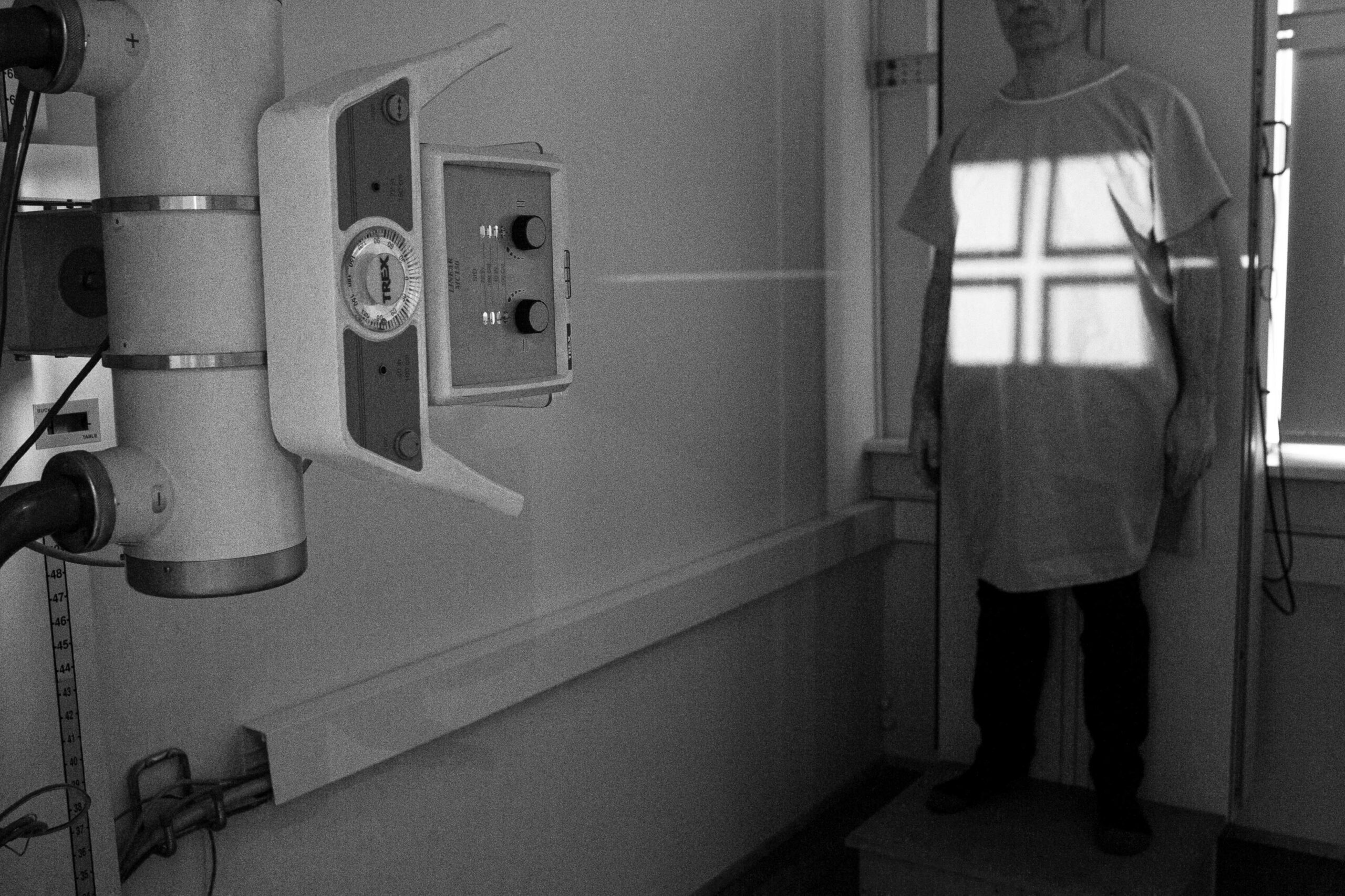
Chiropractors have used X-rays for informational purposes for about one hundred years, from the first years of X-ray imaging. They take different images depending on the information needed and the chiropractic methods being used. Doctors of chiropractic have the education, knowledge and experience to own and operate X-ray equipment and analyze the images. This allows them to obtain exceptional information about the skeletal system and general health of the individual, allowing for better results for the patient.
Why do chiropractors use X-rays in their practice?
- An X-ray supplies a clear image of the skeletal system.
- It allows the doctor to get an accurate count of vertebrae.
- It supplies important information about the position of joints and thereby assists the doctor in adjusting them back into a normal position in the shortest and most specific way possible.
- It also gives valuable information on the general state of joints, possible degeneration, birth defects, arthritis, how joints bear weight, broken bones and other possible underlying trauma to bone structures.
- It may show possible serious disease processes.
X-ray imaging is a relatively safe tool to obtain valuable information about peoples health. Chiropractors who do the Gonstead work use full spine X-rays. Two images are taken. One where the spine can be seen from the side and another one where the spine can be seen from the back.This type of imaging is unique in two ways:1) The whole spine is visible on both films.2) The patient is standing with equal weight on both feet when the films aretaken.Our skeletal system has two major roles. One is to protect our nervous system andthe other one is to bear our weight. By taking the film with the skeletal system in a weightbearing position we are able to obtain information that would be lost if the film was taken with the patient lying down.